Committee on the Histology of Cardiomyopathies: Chairman: B. Maisch
Committee: B. Bültman, S. Factor, H-J Gröne, G. Hufnagel, K. Kawamura, U. Kühl, E.J. Olsen, S. Pankuweit, R.Virmani
Invited Consultants: W. McKenna, P.J. Richardson, G. Thiene, H-Peter Schultheiß, M. Sekiguchi
Committee on Viral Cardiomyopathy: Chairman: B. Maisch
Committee: Ch. Aepinus, K. Aitken, E. Arbustini, L. Archard, C. Baboonian, N. Bowles, S. Broor, G. Hufnagel, R. Kandolf, P. Liu, A. Matsumori, W. McKenna, S. Pankuweit, M. Pauschinger, H-P. Schultheiß, W. Slenczka, M. Sole, K.K. Talwar, J. Towbin, S. Tracy
Invited Consultants: A. Bayes de Luna, J.F. Goodwin, P.J. Richardson
Address for correspondence:
Professor Bernhard Maisch, M.D.
Chairman of the Department of Internal Medicine-Cardiology
Philipps-University Marburg
Baldingerstr. 1, D-35033 Marburg
Germany
Fax: +49-6421-286-8954
Tel: +49-6421-286-6461
WHF Classification and Consensus Conference on the Histo-
and Immunohistopathology of Myocarditis
Marburg, April 28-29, 1997 and on Viral Cardiomyopathy
Marburg, October 3-5, 1997
Introduction
World Health Organisation / International Society and Federation of Cardiology Task Force (ISFC, presently World Heart Federation - WHF) on the Definition and Classification of Cardiomyopathies from 1995 introduced several changes in the definition and classification of heart muscle diseases (1). Since then, the term cardiomyopathy is no longer reserved for the idiopathic forms but can be used interchangeably with the term heart muscle diseases. Right ventricular cardiomyopathy, valvular, hypertensive, ischemic or inflammatory cardiomyopathy have been introduced for the first time as cardiomyopathies.
This new definition of the hemodynamically identified group of dilated cardiomyopathies also comprises INFLAMMATORY CARDIOMYOPATHY. It has been defined as "myocarditis in association with cardiac dysfunction". Idiopathic, autoimmune, and infectious forms of inflammatory cardiomyopathy were recognised.
Within the context of inflammatory cardiomyopathy terms such as active/acute or chronic myocarditis, the association between pericardial disease and myocarditis (perimyocarditis), autoreactive or virally induced myocarditis needed further explanation and diagnostic consensus. Therefore, the WHF Council on Cardiomyopathies formed two expert committees, one with international experts on histopathology and immunohistochemistry, and another one with international experts on the molecular diagnoses of infective or viral cardiomyopathies, which convened in seperate sessions in Marburg, Germany, under the chairmanship of the chairman of the Council on Cardiomyopathies. These experts formulated new definitions of chronic myocarditis and inflammatory dilate cardiomyopathy and on viral cardiomyopathies which are reported here in brief and will be published in extenso soon.
Expert Committee on the histology of dilated inflammatory cardiomyopathy - DCMI
Definition, methodology, and results
The committee defined myocarditis as a process characterised by an inflammatory infiltrate of the myocardium. In acute (active) myocarditis necrosis and/or degeneration of adjacent myocytes is required, whereas in chronic myocarditis necrosis is not an obligatory feature by definition (Figure 1). When referring to the Dallas criteria the term acute myocarditis corresponds to active myocarditis, chronic myocarditis may be defined as comprising borderline or healing myocarditis.
The inflammatory infiltrate should be subclassified as lymphocytic, eosinophilic, neutrophilic, giant cell, granulomatous, or mixed. The distribution should be classified as focal, confluent, or diffuse, respectively.
The panel has chosen for the definition of myocarditis a minimum of 14 infiltrating leukocytes/mm² preferably T-lymphocytes (CD45ro) or activated T-cells (e.g. CD45ro) + (up to 4 macrophages may be included in this total amount). The total number is more than two standard deviations above the number of leukocytes found in control tissue (2-4). In case of nests of leukocytes (>3 lymphocytes, preferably T-cells) located outside the lumen of a vessel, a focal inflammatory process(myocarditis) is diagnosed. If foci of T-lymphocytes are present, myocarditis can be diagnosed due to the nature of the infiltrate even when the critical number of 14 leukocytes/mm² is not reached. If the focal or diffuse leukocytes are localised in fibrotic areas the process may be termed reparative.
The amount and distribution of fibrosis should be described similarly as no (grade 0), mild (grade 1), moderate (grade 2), or severe (grade 3). Localisation or formation of fibrosis should be outlined as endocardial, replacement, or interstitial. Thus the following terminology was adopted:
First biopsy:
Expert Committee on the definition of viral cardiomyopathy
Since isolation of the virus from swabs or tissue is possible only in the acute phase of infection it is unlikely to succeed with this method in patients with longer lasting diseases or chronic infections. Enteroviruses have therefore been effectively isolated only in pediatric patients. The isolation of the active virus remains a standard procedure but has the disadvantage of being time consuming. It was therefore deliberately excluded in this analysis.
A higher sensitivity was achieved with molecular techniques. It is well documented that molecular techniques e.g. gene amplification are significantly more sensitive than standard histochemical technique for the detection of viral proteins.
Except for HIV, hepatitis C and CMV serological assessment of antiviral antibodies appeared to be of limited diagnostic value with respect to the actual disease status of the patients and for the critical issue if the viral genome is present in the myocardium.
Definition, methodology, and results
This WHF expert panel reached a consensus on current diagnostic approaches to viral heart disease by means of an international, multicenter and blinded interlaboratory study. Detection of viral nucleic acid in the myocardium was regarded as indicative of virus infection of the heart. The PCR technique was selected for this study because of its rapidity, wide availability, high sensitivity, and specificity. In situ hybridization, not carried out in this trial, offers near equivalent sensitivity to PCR combined with localization of the virus on the cellular level, with the draw back of the lack of rapidity. PCR primers can be designed to be specific to amplify any member of a particular virus group. The individual agent group can then be identified by direct sequencing of the PCR-product.
The highest sensitivity and reproducibility for the detection of enteroviral genomes were achieved with frozen tissue (100%) in 5 out of 9 centers. Reverse transcription (RT) - PCR of enterovirus RNA from fixed embedded tissue was less reliable, probably with false negatives and the frequent failure to amplify sequences from the processed mRNA of control house keeping genes. Detection of enterovirus sequences in formalin-fixed samples was less convincing. The incidence of hepatitis C virus in formalin fixed tissue (15%) was remarkable. PCR for the genomic sequences of DNA viruses in formalin fixed tissue is less critical and adeno-(12.5%-22.5%), cytomegalo- (5%) and Epstein-Barr virus (2.5%) could also be detected in formalin fixed tissue.
As entero- and adenoviruses are probably the most common agents of viral heart muscle disease, reverse transcription-PCR is required to amplify these viral genomic RNA sequences. The centers experience was that fresh frozen tissue (biopsy) is the material of choice, giving high sensitivity and specificity of detection. Nested PCR seems desirable to detect a low copy number of enteroviral RNA in chronic disease, but single step PCR with Southern blot gave equivalent positive results in the tissue samples and dilution experiments in this study.
An advantage of PCR over slot-blot or in situ hybridization techniques is that where various members of a virus group may be aetiologic, group-specific primers can be used to amplify viral sequences and the particular agent identified subsequently by direct nucleotide sequencing of the PCR product.
Conclusions and recommendations
The WHF expert committee on the histology of inflammatory cardiomyopathy introduced chronic myocarditis as a histologically defined independent category (presence of a diffuse or focal leucocytic infiltrate or foci of lymphocytes associated with the presence of myocellular hypertrophy, focal or diffuse interstitial, replacement and/or perivascular fibrosis and non obligatory microvascular changes) for dilated cardiomyopathies. The presence of chronic inflammatory cells (e.g. lymphocytes, monocytes or macrophages) defined by histology and/or immunohistochemistry in association with the cardiomyopathic changes define chronic myocarditis or dilated cardiomyopathy with inflammation - DCMI.
Chronic myocarditis was defined interchangeably with dilated cardiomyopathy with inflammation or inflammatory cardiomyopathy - DCMI. Considerable variability in the histological diagnosis of chronic myocarditis can often be resolved by immunostaining, which could be helpful in providing more uniform and quantitative criteria for the diagnosis of myocarditis and dilated cardiomyopathy and for the present and future treatment trials.
On the basis of the interlaboraty analysis the second WHF expert panel on viral cardiomyopathies has given for the first time a reliable comparative analysis of cardiac tissue samples infected in part with cardiotropic viruses. The high reproducibility of results for enterovirus positive samples in frozen material by the methods outlined here is an important step for the standardization of diagnostic criteria on viral or inflammatory cardiomyopathy. It has also clearly demonstrated that hepatitis C is an important RNA virus to be considered, as are DNA viruses e.g. adeno- and cytomegalovirus. These findings have to be taken into account on future diagnostic and therapeutic studies in the field of dilated cardiomyopathies and will be published in extenso shortly.
References
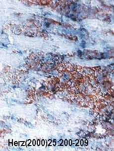
Cryostat sections demonstrating CD4 positive
lymphocytes in chronic myocarditis (magnification
80x). Figure from
Bernhard Maisch, Irene Portig, Arsen Ristic, Günther
Hufnagel & Sabine Pankuweit: Definition of Inflammatory Cardiomyopathy
(Myocarditis): On the Way to Consensus. A Status Report. The article is
available in PDF format: Herz
25 (2000) 3, 200-209